Welcome to the Adie Lab
Our research program consists of three main areas. We seek to (1) advance optical imaging science and technology, (2) develop approaches to enable 4D micromechanical imaging of cell-matrix interactions, and 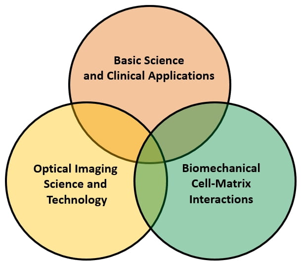 (3) utilize our new imaging approaches for basic science investigations, then translate these approaches and potential discoveries into clinical settings. Our parent imaging modality is optical coherence tomography (OCT), but we also seek to integrate OCT with ultrasound and confocal/multiphoton microscopy. An overview of our three main research areas are below, with further details at our Research page.
(1) Computational imaging and adaptive optics. We are developing novel approaches to acquire and reconstruct OCT images, in order to extend the speed and imaging depth limits of optical microscopy. We introduced ‘hybrid adaptive optics’ (hyAO), an imaging approach that integrates computational and hardware adaptive optics, and have used it to enhance volumetric throughput of cellular-resolution OCT, and to suppress the effects of multiple scattering (a key factor limiting imaging depth in optical microscopy). Through collaborative efforts, we are extending hyAO to multimodal imaging, in particular to help adaptive-optics three-photon microscopy (AO-3PM) image faster and deeper.
(2) Optical coherence elastography (OCE) and traction force microscopy (TFM). We are developing new approaches for 4D imaging of biophysical cell-extracellular matrix (ECM) interactions, including cellular traction forces and ECM mechanical properties. These efforts take advantage of the label-free, interferometric, and deep imaging capabilities of OCT, and make use of localized ‘pushing/palpation’ forces of photons or focused ultrasound. Our ‘home-built’ imaging systems and computational methods uniquely support long-term imaging of dynamic cell-ECM interactions during collective cell invasion in tumor spheroid collagen cell cultures. Our work on developing OCE methods based on ‘ultrasound pushing/palpation’ is to enable mechanical microscopy of the tumor microenvironment in vivo.
(3) Basic science and clinical applications. We leverage the advantages of our custom-designed and built OCT and multimodal imaging systems to better understand the development and progression of disease. Of particular interest is the application of our new imaging techniques to the field of Cell Mechanics and Mechanobiology. We also aim to translate our new imaging capabilities and the discoveries that they may enable to impact the diagnosis or treatment of disease. These efforts benefit greatly from multidisciplinary collaborations with other researchers at Cornell (main Ithaca campus as well as Weill Cornell Medicine in New York City).
If you think your interests and background could be a good match for our research, check out our Positions page.